Computer vision helps detect abnormal blood cells in sickle cell disease
News Sanquin Researcher Tess Afanasyeva ran a pilot study using computer vision to analyze the shape of red blood cells in sickle cell disease, a hereditary blood disorder. Understanding these cell shapes better helps doctors diagnose the disease and track how well treatments are working in real time.
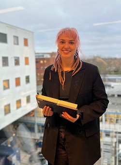
Healthy red blood cells are round and flexible, like a doughnut without a hole. In sickle cell disease, some cells take on a curved "sickle" shape, but many other irregular shapes exist too —some even look like holly leaves, the kind seen in Christmas decorations. These different shapes can tell doctors a lot about a patient’s health.
 New imaging technologies can take pictures of red blood cells in a single drop of blood, but each drop contains thousands of cells—too many for doctors to analyze by eye. That’s where computer vision comes in. Tess trained a computer model on 140,000 images to automatically recognize different cell shapes. Unlike previous studies that relied on machines costing up to half a million euros, Tess developed an AI method that works on more affordable systems. Her ultimate goal is to bring this technology to hospitals in developing countries, where sickle cell disease is most common.
Now, Tess is expanding the project to detect other blood disorders, like spherocytosis and elliptocytosis, using artificial intelligence. This study was made possible with support from the Sanquin Research Fund. This also helped her team buy a powerful NVIDIA graphics card, which will continue to be used for future research at Sanquin.